
Fig. 6
- ID
- ZDB-FIG-150211-23
- Publication
- Paulus et al., 2014 - Loss of Optineurin In Vivo Results in Elevated Cell Death and Alters Axonal Trafficking Dynamics
- Other Figures
- All Figure Page
- Back to All Figure Page
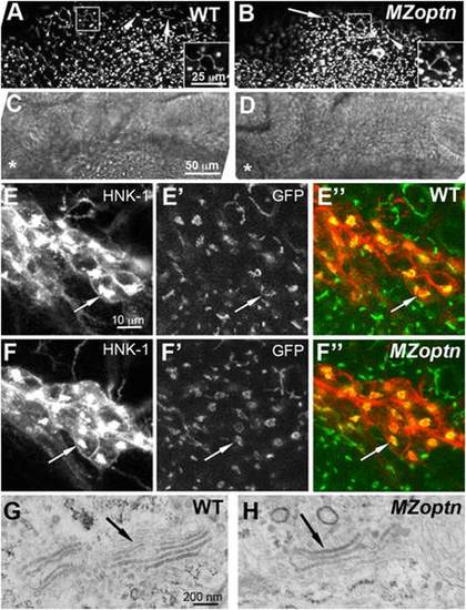
Golgi structure and axon guidance are normal in optn embryos. A–F. Compressed z-stacks of 25 hpf WT (A, C, E) or MZoptn (B, D, F) embryos labeled with man2a-EGFP (A, B, E′, E′′, F′, F′′) or α-HNK-1 (E, F) or brightfield images (C, D). Insets in A, B are higher magnification views of the boxed regions. C, D are brightfield images of the same regions shown in A, B. Anterior is to the left and dorsal is up. A–D. Man2a-GFP labeled Golgi are normal in the midbrain region of MZoptn embryos as seen in both the filamentous Golgi of epithelial cells (arrows) and compact Golgi of neuroepithelial cells (arrowheads). Asterisk indicates location of the eye. E, F. Co-labeling of the sensory trigeminal nucleus with HNK-1 (E, F), man2a-GFP (E′, F′) or the merge of the two channels (E′′, F′′). The Golgi looks normal in MZoptn embryos (arrows). G, H. TEM images of the Golgi in epithelial cells of the head in 25 hpf embryos shows no difference in structure between WT (G) and MZoptn (H) embryos (arrows). |
| Fish: | |
|---|---|
| Observed In: | |
| Stage: | Prim-5 |