Fig. 3
- ID
- ZDB-FIG-150209-1
- Publication
- Housley et al., 2014 - Translational profiling through biotinylation of tagged ribosomes in zebrafish
- Other Figures
- All Figure Page
- Back to All Figure Page
|
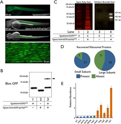
Skeletal muscle-specific translating ribosome purification in zebrafish embryos. (A) Fluorescence image of Tg(ubb:birA-EGFP) expression (upper panel) at 48hpf. Merged fluorescence and bright-field images of Tg(actc1b:Avi-EGFP-rpl10a) expression at 48hpf in the middle two panels show lateral and dorsal views. The bottom panel is a confocal image of Tg(actc1b:Avi-EGFP-rpl10a) expression in skeletal muscle. Anterior is towards the left; dorsal towards the top. (B) Streptavidin shift assay with lysates from 15 embryos from Tg(ubb:birA-EGFP)s972 crossed with wild type (lane 1), from Tg(actc1b:Avi-EGFP-rpl10a)s973 crossed with wild type (lane 2), or from Tg(ubb:birA-EGFP)s972 crossed with Tg(actc1b:Avi-EGFP-rpl10a)s973 (lane 3). Arrowhead indicates Avi-EGFP-Rpl10a bound with free streptavidin. Protein from four larvae at 4dpf was applied to each lane. (C) Eluates from the skeletal muscle TRAP performed on 200 embryos (36hpf) from Tg(ubb:birA-EGFP)s972 crossed with Tg(actc1b:Avi-EGFP-rpl10a)s973. Eluates were separated by SDS-PAGE and stained with Sypro Ruby protein stain (lane 1 is from sibling controls, whereas lane 2 is from embryos expressing both transgenes). Lanes 3 (sibling controls) and 4 (TRAP embryos) depict an ethidium bromide stained agarose gel following Trizol extraction. (D) Illustration of ribosomal proteins with at least one peptide recovered by mass spectrometry analysis of skeletal muscle TRAP eluates. The full list is provided in supplementary material Table S1. (E) qPCR of skeletal muscle and non-skeletal muscle genes from reverse transcribed mRNA purified by TRAP or from whole lysate (input) from 48hpf embryos generated by crossing Tg(ubb:birA-EGFP)s972 to Tg(actc1b:Avi-EGFP-rpl10a)s973. The data are shown as relative abundance of purified mRNA divided by total input mRNA (i.e. TRAP/input) normalized to act1b. Error bars represent s.e.m. from three independent experiments. |
| Gene: | |
|---|---|
| Fish: | |
| Anatomical Terms: | |
| Stage: | Long-pec |

