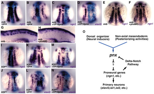
Pnx can promote primary neurogenesis independently of Delta-Notch signaling. (A-H) Ten picograms of pnx and β-galactosidase RNA, 20 pg of VP16pnx and β-galactosidase RNA, or 2 ng of pnx MO (recognizing the translational initiation site) were injected into embryos of crossed mib mutants, fixed at the three-somite stage, and stained with ngn1, elavl3 and islet2 (A-H). The blue region stained with X-gal contained the injected RNA (A-D). The mib homozygous mutant embryos were detected by an increase in Rohon-Beard neurons. Misexpression of pnx in mib expanded the ngn1- (A) and elavl3-expressing stripes (B) but did not change the density of those cells within the stripes. Expression of VP16-Pnx (C,D) or injection of pnx MO (E,F) into the mib mutant embryos reduced the numbers of ngn1 (C,E,F) and elavl3-expressing cells (D) at the three-somite stage. islet2-expressing primary motoneurons were reduced in the pnx MO-injected mib mutant embryos (H). (I-N) One hundred picograms of XDlstu RNA and β-galactosidase RNA were co-injected with 10 pg of pnx RNA or 20 pg VP16pnx RNA into the wild-type embryos, and the expression of ngn1 and elavl3 at the three-somite stage was detected. Expression of XDlstu increased the density of ngn1- (I) and elavl3-expressing cells (J), and co-expression of Pnx expanded the ngn1- (K) and elavl3-expressing (L) stripes without affecting the densities of ngn1- and elavl3-positive cells. Co-expression of VP16-Pnx reduced the numbers of ngn1- (M) and elavl3-expressing cells (N). (A-F,I-N) Dorsal views. (G,H) Lateral views in the spinal cord region. (O) Model for the role of pnx in posterior neurogenesis.
|
