Fig. 1
- ID
- ZDB-FIG-140804-30
- Publication
- Beretta et al., 2013 - The ventral habenulae of zebrafish develop in prosomere 2 dependent on Tcf7l2 function
- Other Figures
- All Figure Page
- Back to All Figure Page
|
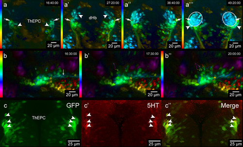
A subpopulation of thalamic-epithalamic early projecting cluster (ThEPC) cells may contribute to the habenulae. (a-a”’) Color code MIP, dorsal views with anterior to the top of the habenula area in living Et(1.0otpa:mmGFP)hd1 embryos at 46 hpf, 57 hpf, 68 hpf and 79 hpf (left to right; see Additional file 1: Movie S1). LUT (look up table) shows the z color code with a z-depth value of 300 μm. Arrows indicate the most anterior ThEPC cells, which appear to end up in the lateral part of the habenulae. (a) Arrowheads indicate the left and right ThEPCs. (a′) Arrowheads indicate the left and the right dHb domain. (a′′′) Arrowheads highlight ThEPC cells, which remain outside the habenulae (circles). (b-b′′) Spectrum MIP, dorsal views with anterior to the left of left-sided ThEPC cells at 48 hpf, 49 hpf and 52 hpf (see Additional file 2: Movie S2). LUT (look up table) shows the z spectrum with a z-depth value of 200 μm. Dots and lines indicate examples of manually tracked dividing cells. (b) Arrow indicates ThEPC axons. (c-c′′) Dorsal view of the thalamic area, MIP, anterior to the top, of a 48 hpf Et(1.0otpa:mmGFP)hd1 embryo stained for 5HT. From left to right: GFP, red and merged channels. Arrowheads highlight some co-labeled GFP/5HT positive neurons. The gamma was adjusted to a value of 0.80 (b,c). dHb, dorsal habenula; ThEPC, thalamic-epithalamic early projection cluster; MIP, maximum intensity projection. |

