Fig. S1
- ID
- ZDB-FIG-140728-19
- Publication
- Acosta et al., 2014 - Mutant Human FUS Is Ubiquitously Mislocalized and Generates Persistent Stress Granules in Primary Cultured Transgenic Zebrafish Cells
- Other Figures
- All Figure Page
- Back to All Figure Page
|
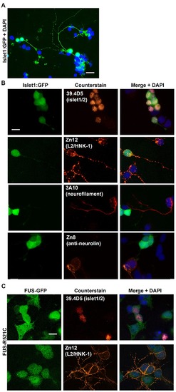
Zebrafish cell culturing protocol supports the growth and differentiation of motor neurons. (A) Cell cultures from transgenic zebrafish embryos expressing GFP under the motor neuron promoter islet-1 (islet1: GFP) demonstrated that motor neurons represented <10% of the cells in culture and exhibited extensive differentiation with axonal growth and branching (arrow). (B) Zebrafish neural cell-associated antibodies obtained from the Developmental Studies Hybridoma Bank (University of Iowa) were used: 39.4D5 [anti-islet-1/2] – primary motor neuron-specific transcription factor; Zn12 [anti-L2/HNK-1] – neural cell adhesion molecule (labels many different neural subtypes); 3A10 [anti-neurofilament] - derived from a neurofilament-associated antigen and labels a subset of hindbrain spinal cord projecting neurons such as Mauthner neurons (Brand et al. 1996) but appears not to label islet 1/2 expressing motor neurons; Zn8 [anti-neurolin] - expressed by secondary but not primary motoneurons during zebrafish development. This labeling demonstrated a mix of different neural subtypes in the cultures. (C) FUS-GFP was expressed in the cell soma of motor neurons and was not extensively transported into neurites. Scale bar = 20 μm. |

