
Fig. 4
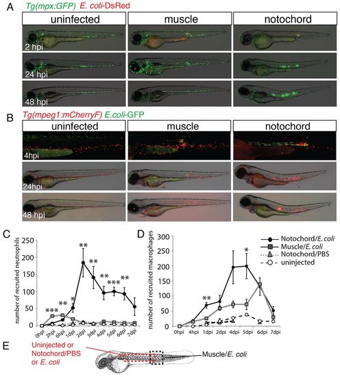
Neutrophils and macrophages are recruited around the infected notochord. (A) Tg(mpx:GFP) larvae were either left uninfected or infected with DsRed-expressing E. coli (red) in the muscle or notochord at 48 hpf. Neutrophil recruitment (green) was imaged repeatedly in individual larvae at the indicated timepoints. (B) A similar experiment to that shown in A was performed in Tg(mpeg1:mCherryF) larvae that were either uninfected or infected with GFP-expressing E. coli (green) in muscle or the notochord at 48 hpf. (C,D) Corresponding counts of neutrophils (C) and macrophages (D) over the course of 1 week post-infection. Results are expressed as the mean number of cells±s.e.m., five to nine fish were examined per timepoint, *P<0.05, **P<0.005 and ***P<0.001. (E) The dashed boxes in the diagram represent the zone where counting was performed. |