
Fig. 7
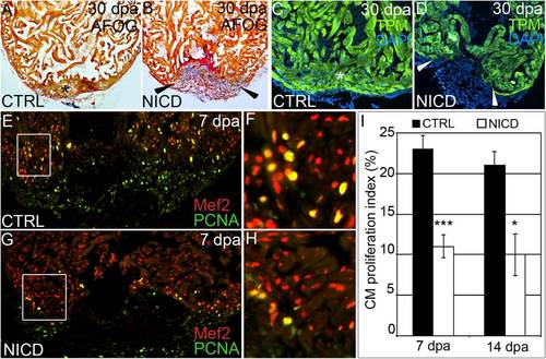
Hyperactivation of Notch signaling impairs cardiomyocyte proliferation and heart regeneration. (A–D) Representative cardiac sections from heat shocked control (CTRL) (A and C) and Tg(hsp70:Gal4); Tg(UAS:NICD) (B and D) animals at 30 dpa stained with AFOG (A and B) or an antibody recognizing the myocardial marker TPM (C and D). Whereas heat shocked CTRL animals regenerated myocardium (asterisks in A and C), Tg(hsp70:Gal4); Tg(UAS:NICD) hearts exhibited impaired regeneration (arrowheads in B and D), residual fibrin (red in B), and collagen deposition (blue in B). In the CTRL group, >25 hearts were analyzed and all showed significant myocardial regeneration. In the Tg(hsp70:Gal4); Tg(UAS:NICD) group, 18 hearts were analyzed and 14 showed a significant myocardial deficit, residual fibrin, and collagen deposition. (E–H) Representative cardiac sections of heat shocked CTRL (E and F) and Tg(hsp70:NICD) (G and H) animals at 7 dpa double stained with antibodies that recognize cardiomyocyte nuclei (Mef2+) and nuclei undergoing DNA replication (PCNA+). Boxed regions in E and G are shown at higher zoom in F and H. (I) The percentages of cardiomyocyte nuclei undergoing DNA replication were quantified along the wound edge at 7 and 14 dpa and expressed as mean proliferation indices ± 1 SD, ***P < 0.001; *P < 0.05. In the control group, seven and eight hearts were analyzed at 7 and 14 dpa, respectively. In the Tg(hsp70:NICD) group, three and four hearts were analyzed at 7 and 14 dpa, respectively. |