
FIGURE
Fig. S3
- ID
- ZDB-FIG-140422-49
- Publication
- Holly et al., 2014 - Sfrp1a and Sfrp5 function as positive regulators of Wnt and BMP signaling during early retinal development
- Other Figures
- All Figure Page
- Back to All Figure Page
Fig. S3
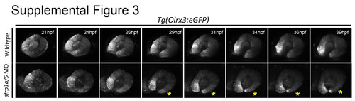
Fig. S3. Live embryo analysis of sfrp1a/sfrp5 depletion during retinal morphogenesis. Time lapse live confocal imaging of the Tg(Olrx3:GFP) line was employed to visualize retinal morphogenesis. Images are maximum projections of 30 slices collected every 3 μm at 10 minute intervals. Compared to WT, sfrp1a/5 morpholino injected embryos display thinning of the ventral retina as development proceeds post-24 hpf (frames 26 hpf to 39 hpf). Thinning retinal tissue is indicated with a yellow asterisk. |
Expression Data
Expression Detail
Antibody Labeling
Phenotype Data
Phenotype Detail
Acknowledgments
This image is the copyrighted work of the attributed author or publisher, and
ZFIN has permission only to display this image to its users.
Additional permissions should be obtained from the applicable author or publisher of the image.
Reprinted from Developmental Biology, 388(2), Holly, V.L., Widen, S.A., Famulski, J.K., and Waskiewicz, A.J., Sfrp1a and Sfrp5 function as positive regulators of Wnt and BMP signaling during early retinal development, 192-204, Copyright (2014) with permission from Elsevier. Full text @ Dev. Biol.