
FIGURE
Fig. S2
Fig. S2
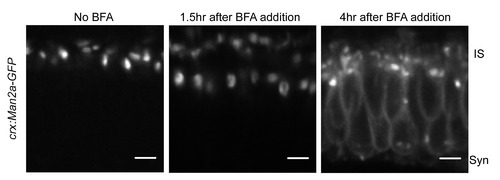
Man2a-GFP marks medial Golgi structures. We confirmed that our Golgi marker behaved in the same manner as endogenous Golgi proteins by treating 3 dpf Tg(crx:Man2a-GFP) zebrafish larvae with 2 μM Brefeldin A followed by live imaging. After a 4 hour incubation in BFA, the GFP signal was present in the ER and fragmented Golgi structures. |
Expression Data
Expression Detail
Antibody Labeling
Phenotype Data
Phenotype Detail
Acknowledgments
This image is the copyrighted work of the attributed author or publisher, and
ZFIN has permission only to display this image to its users.
Additional permissions should be obtained from the applicable author or publisher of the image.
Full text @ PLoS One