
FIGURE
Fig. S4
Fig. S4
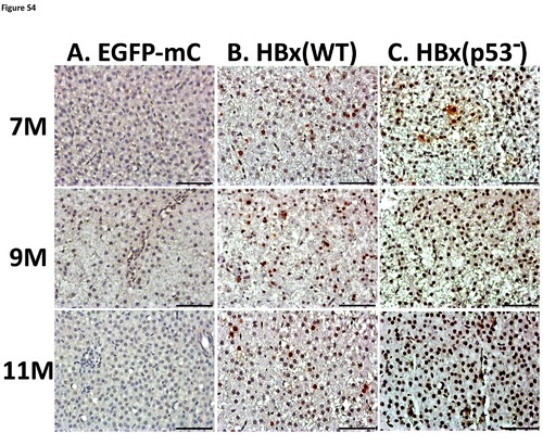
Representative images of PCNA staining in HBx overexpression transgenic fish. (A) EGFP-mCherry transgenic fish, (B) HBx overexpression in wild-type, or (C) HBx overexpression in p53 mutant transgenic fish stained with PCNA antibody 7, 9 and 11 months (x 400).Scale bars: 50μm. |
Expression Data
Expression Detail
Antibody Labeling
Phenotype Data
Phenotype Detail
Acknowledgments
This image is the copyrighted work of the attributed author or publisher, and
ZFIN has permission only to display this image to its users.
Additional permissions should be obtained from the applicable author or publisher of the image.
Full text @ PLoS One