
Fig. S2
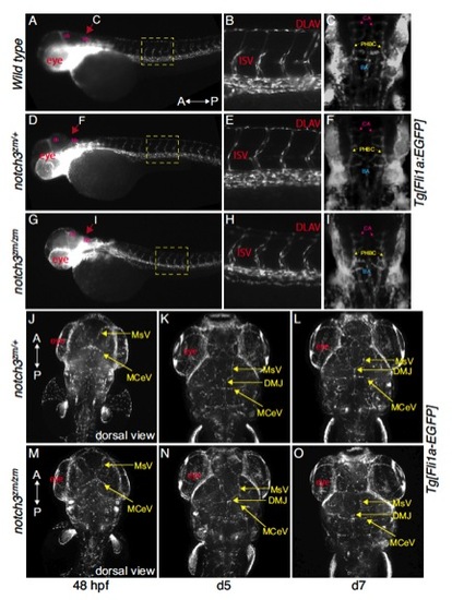
Tg[fli1a:EGFP] labelled vasculature of A,B,C) wild-type, D,E,F) notch3zm/+, G,H,I) notch3zm/zm were indistinguishable. A-I) 52hpf. A,B,D,E,G,H) Lateral views; max projection images. Dorsal is to the top and the Anterior (A) and posterior (P) axis is as indicated. A,D,G) The yellow dashed boxes correspond to the regions shown in the higher magnifications in B,E,H. C,F,I and J-O) Dorsal views; max projection images of Tg[fli1a:EGFP] labelled vasculature system of C) wild type F, J-L) notch3zm/+ heterozygotes and I, M-O) notch3zm/zm mutants. Vessel pattern at the anatomical level is intact in notch3zm/zm mutants (48hpf=9 hets and 6 mutants; day 5=2 hets, 3 mutants; day 7 = 3 WT, 6 hets, 2 mutants). The orientation of the Anterior (A) and posterior (P) axis is indicated. DLAV indicates the Dorsal Longitudinal Anastomotic Vessel; ISV denotes the InterSegmental Vessels; CA indicates the central artery; PHBC denotes the primordial hindbrain channel; BA indicates the Basilar artery; MsV indicates the Mesencephalic Vein; DMJ denotes the Dorsal Midline Junction; MCeV indicates the Mid-Cerebral Vein. |