
Fig. S2
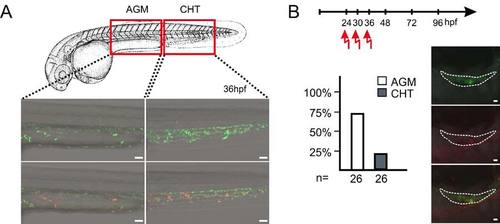
Time and place of origin of thymus-settling precursors (A) Green fluorescent hematopoietic cells of fish transgenic for ikaros:tdEosFP were photoconverted in the indicated regions of the embryo (AGM, aorta-gonad-mesonephros; CHT, caudal hematopoietic tissue) at various time points (here shown for 36hpf); n=26 embryos for each time-point and region. Scale bars, 50μm. |
Reprinted from Immunity, 36(2), Hess, I., and Boehm, T., Intravital Imaging of Thymopoiesis Reveals Dynamic Lympho-Epithelial Interactions, 298-309, Copyright (2012) with permission from Elsevier. Full text @ Immunity