Fig. 6
- ID
- ZDB-FIG-130910-25
- Publication
- Kim et al., 2013 - Multi-organ Abnormalities and mTORC1 Activation in Zebrafish Model of Multiple Acyl-CoA Dehydrogenase Deficiency
- Other Figures
- All Figure Page
- Back to All Figure Page
|
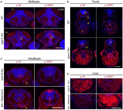
Tissue dependent regulation of mTORC1 activation in dxavu463 mutant zebrafish. Anti-phospho-S6 (left panels) and anti-phospho-4E-BP1 (right panels) antibodies were used to assess mTORC1 kinase activity. (A) WT brain (top) and dxa brain (bottom) at 8 dpf. Arrows indicate p-S6 and phospho-4E-BP1 positive cells in neural progenitors of the brain. P-S6 and p-4E-BP1 are also detected in the superficial pial cells of the mutant brain. (B) Sections of trunk regions in WT (top) and dxa (bottom) at 8 dpf. Phospho-S6 was detected in the central canal and phospho-4E-BP1 positive cells were found central canal as well as midline cells (yellow arrow) in dxa mutant zebrafish. Asterisks indicates central canal of hindbrain. 300 nM of rapamycin was used from 5 dpf to 8 dpf to treat dxa mutant zebrafish in C and D. (C) Hindbrain regions of type III mutant at 8 dpf. Phospho-S6 and phospho-4E-BP1 staining was again detected in central canal (*) and pial cell sheath (P) in both control and rapamycin treated dxa mutants. DAPI (blue) was used for nuclei staining. (D) Liver regions of same sections seen in (C) with marked suppression of phospho-S6 but a relative increase in phsohp-4E-BP1 levels. P, pial cell sheath; HB, hindbrain; K, kidney; L, liver. Scale bar = 100 μm. |
| Antibodies: | |
|---|---|
| Fish: | |
| Anatomical Terms: | |
| Stage: | Days 7-13 |

