
Fig. 1
- ID
- ZDB-FIG-130826-50
- Publication
- Chapman et al., 2013 - Axonal Transport Defects in a Mitofusin 2 Loss of Function Model of Charcot-Marie-Tooth Disease in Zebrafish
- Other Figures
- All Figure Page
- Back to All Figure Page
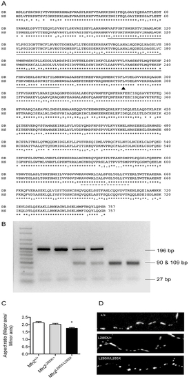
A zebrafish MFN2 mutant. (A) CLUSTAL alignment of zebrafish (Danio rerio: DR) and human (Homo sapiens: HS) Mfn2 protein sequences (Ensembl). The solid and dashed lines indicate the GTPase and transmembrane domains respectively. The site of the ENU induced point mutation (L285) is indicated with an arrowhead. (B) The ENU induced mutation creates a novel restriction enzyme site for MseI, allowing genotyping using PCR followed by MseI digest. The agarose gel shows, from left to right: Hyperladder V (Bioline), two wild type embryos (MFN2+/+), two mutation carriers (MFN2L285X/+), and two homozygotes (MFN2L285X/L285X). (C) Mitochondria in cultured MFN2L285X/L285X neurons have a significantly reduced aspect ratio (p<0.01, Kruskal-Wallis with Dunn’s multiple comparisons test). (D) Representative images of axonal mitochondria stained with Mitotracker Red in cultured neurons of the indicated genotypes. |
| Fish: | |
|---|---|
| Observed In: | |
| Stage: | Prim-5 |