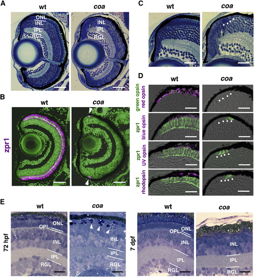
Photoreceptors Undergo Apoptosis in the Zebrafish coa Mutant (A) Plastic sections of WT and coa mutant retinas at 6 dpf. ONL is absent in the coa mutant. (B) Zpr1 antibody labeling of 6 dpf WT and coa mutant retinas. All nuclei are counterstained with SYTOX Green (green). Arrowheads indicate zpr1 expression (magenta) in the CMZ of the coa mutant. (C) Plastic sections of CMZ of 6 dpf WT and coa mutant retinas. White dots indicate morphologically distinct ONL in the coa mutant. (D) Labeling of 6 dpf WT and coa mutant retinas with anti-red, blue, UV opsins, or rhodopsin antibodies (magenta); anti-green opsin antibody (green); and zpr1 (green) antibody. (E) Semithin sections of WT and coa mutant retinas at 72 hpf and 7 dpf. At 72 hpf, pyknotic nuclei were observed in the ONL in the coa mutant (arrowheads). At 7 dpf, ONL was absent in the coa mutant. INL, inner nuclear layer; IPL, inner plexiform layer; OPL, outer plexiform layer; RGL, retinal ganglion cell layer. Scale bars, 50 μm (A and B) and 20 μm (C–E). See also Figure S1.
|
