
FIGURE
Fig. 3
- ID
- ZDB-FIG-130808-12
- Publication
- Hayes et al., 2013 - Expression of glycosaminoglycan epitopes during zebrafish skeletogenesis
- Other Figures
- All Figure Page
- Back to All Figure Page
Fig. 3
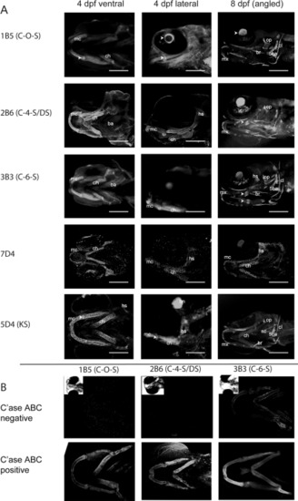
Chondroitin and keratan sulfate expression in the developing jaw. Confocal reconstructions showing comparative regions of the developing jaw of 8-day zebrafish larvae labelled with antibodies towards distinct chondroitin and keratan sulfate epitopes (refer to Table 2 for antibody specificities). In all images larvae are presented with anterior facing to the left. Arrowheads point to ossifying regions of the ceratohyal. ch, ceratohyal; mx, maxilla; mc, Meckel′s cartilage; pq, palatoquadrate. |
Expression Data
| Antibodies: | |
|---|---|
| Fish: | |
| Anatomical Terms: | |
| Stage: | Days 7-13 |
Expression Detail
Antibody Labeling
Phenotype Data
Phenotype Detail
Acknowledgments
This image is the copyrighted work of the attributed author or publisher, and
ZFIN has permission only to display this image to its users.
Additional permissions should be obtained from the applicable author or publisher of the image.
Full text @ Dev. Dyn.