
Fig. 2
- ID
- ZDB-FIG-130722-3
- Publication
- Tseng et al., 2011 - An evolutionarily conserved kernel of gata5, gata6, otx2 and prdm1a operates in the formation of endoderm in zebrafish
- Other Figures
- All Figure Page
- Back to All Figure Page
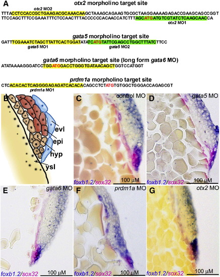
Morpholino and phenotypic characterization of morphants for germ layer progenitor cell fate specification. (A) The nucleotide sequence surrounding the translation initiation site for gata5, gata6, otx2 and prdm1a, with the MO target site highlighted. (B) Illustration of section to demonstrate the cells′ lineage. (C–G) Section double in situ hybridization with ectoderm marker (foxb1.2 shown in blue) and endoderm marker (sox32 indicated by pink) for the 5 hpf morphants injected by the control MO (C), gata5 MO (D), gata6 MO (E), prdm1a MO (F) and otx2 MO (G). MO shown in the right upper corner and scale bar indicated with a black line in the right lower corner. |
| Genes: | |
|---|---|
| Fish: | |
| Knockdown Reagents: | |
| Anatomical Terms: | |
| Stage: | 30%-epiboly |
Reprinted from Developmental Biology, 357(2), Tseng, W.F., Jang, T.H., Huang, C.B., and Yuh, C.H., An evolutionarily conserved kernel of gata5, gata6, otx2 and prdm1a operates in the formation of endoderm in zebrafish, 541-57, Copyright (2011) with permission from Elsevier. Full text @ Dev. Biol.