|
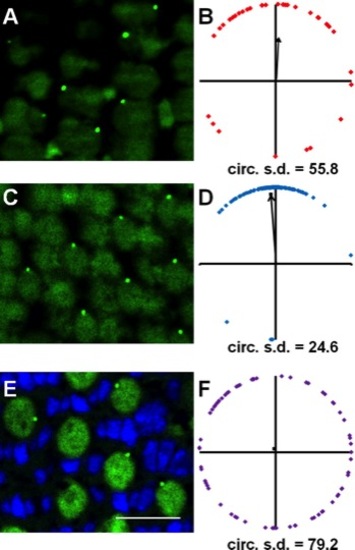
Centrin-GFP labels basal bodies in the retinas of adult Tg(XlRho:gap43-CFP)ucd1 transgenic zebrafish. Sample portions of fields of cells at the depths of the basal bodies of red-/green-sensitive cones (A), blue-sensitive cones (C), and UV-sensitive cones (E). GFP fluorescence localized to basal bodies (green), and nuclei were counterstained with DAPI (blue). Autofluorescence (green) from 488-nm excitation shows inner and outer segments. B,D,F: Graphs of the positions of basal bodies from fields A,C,E in which the positions of all the basal bodies of red-/green-, blue-, or UV-sensitive cones were plotted around a unit circle (red, blue, or purple lozenges, respectively), and the mean vector is indicated (arrow). Optic nerve is upward in all panels. A magenta-green version of this figure is provided in the Supporting Information. Scale bar = 10 μm.
|