Fig. S5
- ID
- ZDB-FIG-130618-11
- Publication
- Villefranc et al., 2013 - A truncation allele in vascular endothelial growth factor c reveals distinct modes of signaling during lymphatic and vascular development
- Other Figures
- (all 10)
- All Figure Page
- Back to All Figure Page
|
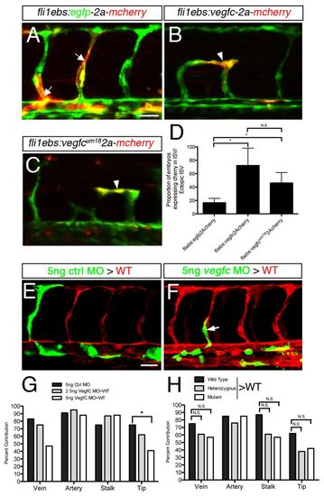
Developmental angiogenesis defects in vegfc-deficient mouse and zebrafish embryos. (A,B) Confocal micrographs of mouse embryos of the indicated genotype at E9.5 stained with endomucin to visualize endothelial cells. (A) Cranial vessels (left) and quantification of branching points (right). (B) ISVs (left) and quantification of added ISV length, between somites 14-16 (right). n=3. (C-F) Confocal micrographs of Tg(fli1a:egfp)y1 wild-type zebrafish embryos at 30 hpf injected with (C) 5 ng control, (D) 2.5 ng Vegfc or (E) 5 ng Vegfc MO. Lateral views, dorsal is up, anterior to the left. (F) Quantification of ISV length in MO-injected Tg(fli1a:egfp)y1 embryos. *P<0.05; N.S., not significant. Scale bars: 100 μm in A,B; 25 μm in C-E. |

