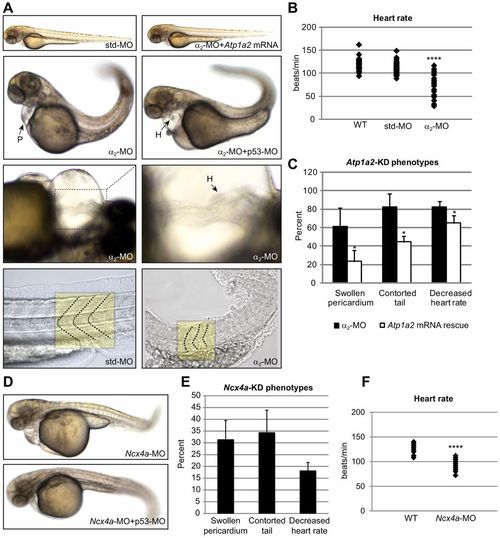
α2-MO-mediated Atp1a2 knockdown affects skeletal and heart muscle. (A) Embryos injected with α2-MO or p53-MO+7alpha;2-MO exhibited a contorted tail, abnormal somite morphology, a swollen pericardium and loss of cardiac laterality. Coinjecting α2-MO with Atp1a2 mRNA rescued knockdown phenotypes demonstrating specificity. Normal and distorted somite borders of std-MO- and α2-MO-injected embryos are indicated by dashed lines in the bottom panel. (B) Heart rates (beats/min) of wild-type (WT), std-MO- and α2-MO-injected embryos at 60 hpf (n = 30) (see supplementary material Movies 1, 2). (C) The penetrance of the observed phenotypes in α2-MO-injected (n = 91) and mRNA-rescued embryos (n = 136), plotted as percentages ± s.d. of embryos displaying the phenotype, relative to total embryo number. (D) Embryos injected with Ncx4a-MO phenocopy the α2-MO-injected embryos. (E) The penetrance of the observed phenotypes in Ncx4a-MO-injected embryos (n = 107), plotted as percentages ± s.d. of embryos displaying the phenotype, relative to total embryo number. (F) Heart rates (beats/min) of WT and bradycardic Ncx4a-MO-injected embryos at 60 hpf (n = 20). *P<0.05, ****P<0.0001.
|
