
Fig. 1
- ID
- ZDB-FIG-130313-14
- Publication
- Hochgreb-Hägele et al., 2013 - A novel FoxD3 gene trap line reveals neural crest precursor movement and a role for FoxD3 in their specification
- Other Figures
- All Figure Page
- Back to All Figure Page
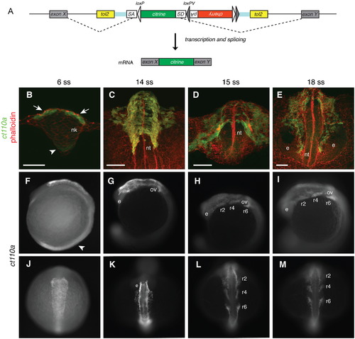
A novel protein trap line allows visualization of neural crest cells from early stages in zebrafish. (A) Schematic diagram of the FlipTrap strategy. The FlipTrap construct contains an internal exon, consisting of citrine sequence lacking start and stop codons (green), flanked by splice acceptor (SA) and donor (SD) sites. Genomic integration of the construct is mediated by Tol2 transposase. When the FlipTrap construct inserts within an intronic region, the endogenous splicing machinery recognizes SA and SD sites, and the citrine sequence is integrated into the mRNA (dashed lines), resulting in an endogenously expressed Citrine-fusion protein. (B–D) Confocal images of ct110a embryos. Transverse sections. C and D are 3D-projections of z-stacks. Embryos counterstained with phalloidin-rhodamine (red) and Citrine signal amplified by immunohistochemistry against GFP after fixation (green). (B) Expression of Citrine is detected in the dorso-lateral cells (white arrow) of the neural keel (nk) at 6-somite stage (ss). Low-level expression can be detected in the floor plate (white arrowhead). (C) Delamination of neural crest cells (NCCs) from the neural tube (nt). Citrine-positive cells distributed on the dorsal aspect of the neural tube at 14ss. (D) Migration of NCCs from the neural tube along stereotyped routes throughout the embryo. (E) At 18ss, cranial NCCs migrate anteriorly to the head, circumventing the eyes (e). Scale bars: 50 μm. (F–M) Expression of Citrine in ct110a embryos. (F–I) Left side view. (J–M) Dorsal view. Citrine signal amplified by GFP immunostaining in fixed embryos. (F and J) Expression of Citrine in NCCs on the dorsal aspect of the anterior neural tube, at the level of the hindbrain. Low expression in the floor plate region (white arrowhead). (G and K) NCCs delaminate and migrate from the neural tube to the head, around the otic vesicles (ov) and eyes (e). (H, I, L and M) NCCs migrate ventrally into the pharyngeal arches in three separate streams along stereotyped routes (r2, r4 and r6). |
| Gene: | |
|---|---|
| Fish: | |
| Anatomical Terms: | |
| Stage Range: | 5-9 somites to 14-19 somites |
Reprinted from Developmental Biology, 374(1), Hochgreb-Hägele, T., and Bronner, M.E., A novel FoxD3 gene trap line reveals neural crest precursor movement and a role for FoxD3 in their specification, 1-11, Copyright (2013) with permission from Elsevier. Full text @ Dev. Biol.