
Fig. 2
- ID
- ZDB-FIG-130218-2
- Publication
- Plazas et al., 2013 - Activity-dependent competition regulates motor neuron axon pathfinding via PlexinA3
- Other Figures
- All Figure Page
- Back to All Figure Page
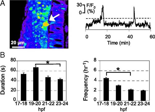
Developing PMN generate spontaneous intracellular Ca2+ waves. (A) PMN (arrow) loaded with CGD in the spinal cord of a zebrafish embryo at 20–21 hpf. Dorsal to the left and caudal at the bottom. Images were captured at 0.2 Hz for 30 min, and fluorescence intensity was normalized to baseline. Fluorescence is displayed on a pseudocolor scale, where purple represents the lowest intensity and red represents the highest intensity. Changes in fluorescence intensity in a PMN are plotted as a function of time. Ca2+ waves were identified as fluorescence transients greater than 20% of ΔF/F0 (dashed lines), more than two times the SD of the baseline, and >20 s in duration, calculated as the width at half-maximum. (B) Duration and frequency of Ca2+ waves in PMN. n = 15–20 neurons from seven to nine embryos for each time period; values are means ± SEM. *P < 0.05. |