
Fig. S1
- ID
- ZDB-FIG-130123-44
- Publication
- Schweitzer et al., 2013 - Sim1a and Arnt2 contribute to hypothalamo-spinal axon guidance by regulating Robo2 activity via a Robo3-dependent mechanism
- Other Figures
- All Figure Page
- Back to All Figure Page
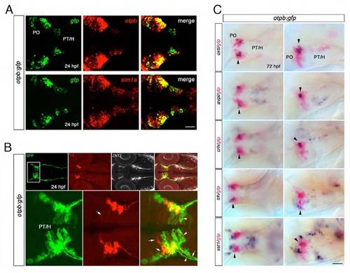
The otpb:gfp reporter line recapitulates many aspects of endogenous otpb expression in the forebrain. (A) Dorsal views of confocal z projections of the forebrain region of otpb:gfp transgenic embryos at 24 hpf, demonstrating co-expression of gfp (green) and otpb (red), as well as co-expression of gfp (green) and sim1a (red) analyzed by double fluorescent in situ hybridization are shown. Scale bar: 50 μm. (B) Dorsal views of otpb:gfp transgenic embryos at 24 hpf co-labeled with anti-GFP (green), anti-TH (red) and anti-ZN12. Dopaminergic TH-positive cell bodies and longitudinal dopaminergic projections are immunoreactive for GFP (arrows). Scale bar: 100 μm. (C) Dorsal (left panel) and lateral views (right panel) of otpb:gfp transgenic embryos at 72 hpf analyzed for co-expression of hypothalamic neurohormones (shown in blue) and gfp (shown in purple) by double in situ hybridization. Oxytocin-like (oxtl), arginine vasopressin-like (avpl), corticotropin releasing hormone (crh), thyrotropin releasing hormone (trh) and somatostatin 1 (sst1) transcripts in the preoptic region all colocalize with gfp expression (see arrowheads). Scale bar: 50 μm. PT, posterior tuberculum; H, hypothalamus; PO, preoptic region. |