|
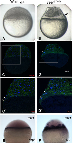
The cea mutant has an enlarged YSL. (A) Wild-type and (B) cea mutant embryos at 4 hpf. Arrow in B indicates the enlarged syncytial layer. (C–D′) Cryosections of wild-type and mutant embryos at 4 hpf, stained with anti-β-catenin antibody (green) and DAPI (blue). C′ and D′ show enlargements of the areas indicated by squares in C and D, respectively. Nuclei (white arrowheads) are not separated by cell membranes in both wild-type and mutant embryos. The clusters of nuclei and surrounding cytoplasm are larger in the cea mutant than the wild-type embryo (compare C′ with D′). (E,F) Whole-mount in situ hybridization of (E) control and (F) cea mutant embryos with the YSL marker, mtx1. The enlarged syncytial layer expresses mtx1 at 6 hpf.
|