
Fig. 2
- ID
- ZDB-FIG-130108-2
- Publication
- Uribe et al., 2012 - Id2a functions to limit Notch pathway activity and thereby influence the transition from proliferation to differentiation of retinoblasts during zebrafish retinogenesis
- Other Figures
- All Figure Page
- Back to All Figure Page
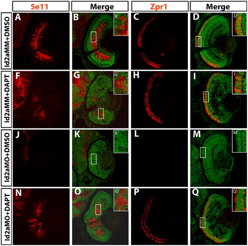
DAPT inhibition of the Notch pathway rescues terminal differentiation in Id2a-deficient retinae. Transverse retinal sections at 72 hpf from Id2aMM-DMSO (A)–(D), Id2aMM-DAPT (F)–(I), Id2aMO-DMSO (J)–(M) and Id2aMO-DAPT (N)–(Q) embryos. Embryos were treated with DMSO or DAPT from 28–72 hpf. Amacrine cells marked by 5e11 (A), (F), (J) and (N) and red/green cones marked by Zpr1 (C), (H), (L) and (P) fail to differentiate in Id2aMO retinas treated with DMSO (J), while in Id2aMO retinas treated with DAPT, both amacrine cells and red/green cones are detected (N) and (P). Merged images show co-staining of retinal marker (red) and nuclei (Sytox-green; green). Dorsal is up in all images. |
| Antibodies: | |
|---|---|
| Fish: | |
| Condition: | |
| Knockdown Reagent: | |
| Anatomical Terms: | |
| Stage: | Protruding-mouth |
| Fish: | |
|---|---|
| Condition: | |
| Knockdown Reagent: | |
| Observed In: | |
| Stage: | Protruding-mouth |
Reprinted from Developmental Biology, 371(2), Uribe, R.A., Kwon, T., Marcotte, E.M., and Gross, J.M., Id2a functions to limit Notch pathway activity and thereby influence the transition from proliferation to differentiation of retinoblasts during zebrafish retinogenesis, 280-292, Copyright (2012) with permission from Elsevier. Full text @ Dev. Biol.