
Fig. 2
- ID
- ZDB-FIG-121217-29
- Publication
- Nguyen-Chi et al., 2012 - Morphogenesis and Cell Fate Determination within the Adaxial Cell Equivalence Group of the Zebrafish Myotome
- Other Figures
- All Figure Page
- Back to All Figure Page
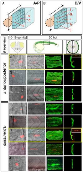
The fate map of the adaxial compartment. The fate of individual adaxial cells were identified using iontophoretic injections of TMRD (red) into individual adaxial cells located at various AP (C-E) and DV (F-J) positions within the three most newly formed somites of 10-15-somite stage embryos (C-J, left panels, dorsal views). The AP (A) and DV (B) position of labeled cells is represented schematically. The fate of cells was then analyzed at 30 hpf (C-J, other panels). (C-J) At 30 hpf, the panels (from the left to the right) represent the position of the labeled fiber in the myotome (lateral view), the expression of sMyHC (green, 3D reconstruction of multiple confocal scans) in lateral view and in cross section, and the expression of Engrailed (green, small upper panels for C and H, lateral view). MPs (C, H) were identified due to their position in the midline and simultaneous expression of Slow MyHC and Eng. |