
Fig. 1
- ID
- ZDB-FIG-120815-65
- Publication
- Hesselson et al., 2011 - Suppression of Ptf1a activity induces acinar-to-endocrine conversion
- Other Figures
- All Figure Page
- Back to All Figure Page
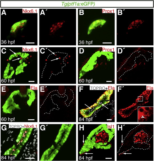
ptf1a Expression Becomes Restricted to Differentiated Acinar Cells during Early Larval Development Embryos express Tg(ptf1a:eGFP) in the pancreatic domain. Expression of pancreatic markers was analyzed by immunofluorescence. Scale bars represent 20 μm. (A–B2) Confocal projections of pancreatic tissue at 36 hours postfertilization (hpf). (C–H2) Confocal sections of pancreatic tissue at 60 (C–E2) and 84 hpf (F–H2). Dotted lines delineate Tg(ptf1a:eGFP)-expressing tissue. (A–B2) Tg(ptf1a:eGFP)-expressing cells initially coexpress Nkx6.1 and Prox1. (C–D2) By 60 hpf, Nkx6.1 (C and C2, arrows) and Prox1 (D and D2) become excluded from the Tg(ptf1a:eGFP) expression domain. (E–F2) Coexpression of Tg(ptf1a:eGFP) with the acinar cell marker Elastase (Ela) is detected by 84 hpf. Elastase-positive granules are detected on the apical surface of acinar cells (F2, arrowhead), adjacent to Tg(ptf1a:eGFP)-negative intrapancreatic cells (F, arrows). (G–H2) The duct marker Nkx6.1 (G and G2) and the endocrine marker Isl1 (H and H2) appear to be restricted to Tg(ptf1a:eGFP)-negative ductal and islet cells respectively by 84 hpf. Isl1 is also expressed in the pancreatic mesenchyme (H and H2, arrows). |