
Fig. 6
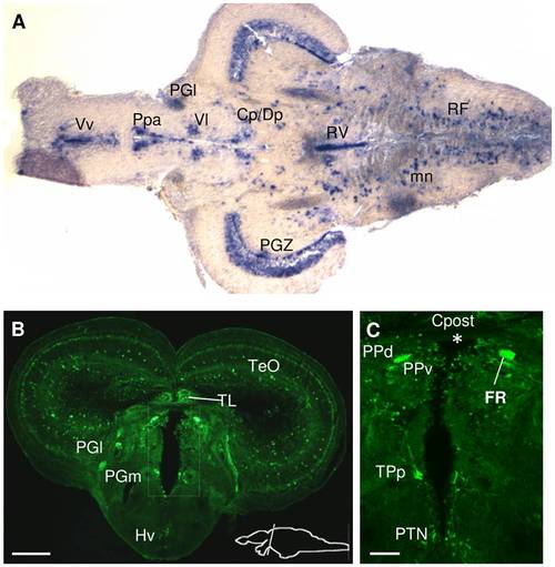
Spon1b expression in thalamic and pretectal regions. A. Horizontal section showing spon1b mRNA-positive nuclei (in situ hybridization): dorsoposterior (DP), centroposterior (CP) and ventrolateral (Vl) thalamic nuclei; anterior parvocellular preoptic (Ppa); Ventral Tel (Vv). B. Coronal section through the thalamus, immunostained for GFP. Medial (PGm) and lateral (PGl) preglomerular nuclei, torus longitudinalis (TL), ventral hypothalamus (Hv), optic tectum (TeO). C. Periventricular region (boxed area in B in adjacent section), showing spon1b-positive cells in the dorsal (PPd) and ventral (PPv) periventricular pretectal nuclei, in the posterior tuberculum (TPp), in the posterior tuberal nucleus (PTN) and in fibers of the fasciculus retroflexus (FR). Note absence of signal in the subcommissural organ (asterisk: SCO region, ventral to posterior commissure, Cpost). Scale bars: A-B: 100 μm; C: 50 μm. |