
Fig. 6
- ID
- ZDB-FIG-120720-61
- Publication
- Okuda et al., 2012 - lyve1 expression reveals novel lymphatic vessels and new mechanisms for lymphatic vessel development in zebrafish
- Other Figures
- All Figure Page
- Back to All Figure Page
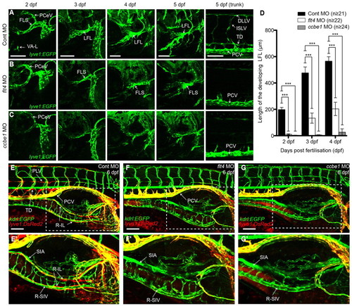
The development of the FL, IL and LL requires flt4 and ccbe1. (A-C) Lateral images of the lyve1:EGFP transgenic, showing altered FL and trunk lymphatics development in flt4 (B) and ccbe1 (C) morpholino-injected embryos compared with control morpholino-injected embryos (A) at 2-5 dpf. (D) Quantification of the length of the developing LFL in control, flt4 and ccbe1 morpholino-injected embryos at 2, 3 and 4 days post fertilisation. Error bars represent 95% confidence intervals. ***P<0.001. (E-G′) Lateral images of the lyve1:DsRed2;kdrl:EGFP transgenic showing lack of IL and LL development in flt4 (F) and ccbe1 (G) morpholino-injected embryos compared with the normal IL and LL development in the control morpholino-injected embryos (E). E′-G′ are higher magnification images of the boxes in E-G. E-G are montage images of two z series stacks. DLLV, dorsal longitudinal lymphatic vessel; FLS, facial lymphatic sprout; ISLV, intersegmental lymphatic vessel; LFL, lateral facial lymphatic; PCeV, posterior cerebral vein; PCV, posterior cardinal vein; PLV, parachordal lymphatic vessel; R-IL, right intestinal lymphatics; R-SIV, right subintestinal vein; SIA, supraintestinal artery; TD, thoracic duct. VA-L, ventral aorta lymphangioblasts. Scale bars: 100 μm. |