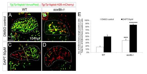
Fig. S4
|
The intrahepatic biliary cells in sox9b mutants are more susceptible to Notch signaling inhibition. (A–D) Confocal images of livers in Tg(Tp1bglob:VenusPest); Tg(Tp1bglob:H2B-mCherry) wild-type (A, C) and sox9b mutant (B, D) larvae treated with DMSO (A–B) or 50 μM DAPT (C–D) from 106 to 154 hpf. DAPT treatment caused an increase in the relative proportion of Tg(Tp1bglob:H2B-mCherry)-single positive cells in all the animals, yet sox9b mutants exhibited a more severe increase compared to wild-type larvae. In DAPT-treated wild-type larvae (C), loss of Notch activity was more prominent in the region proximal to the extrahepatic duct (p, left side of yellow line), whereas the distal biliary cells still maintained Tg(Tp1bglob:VenusPest) expression (d, right side of the yellow line). (A–D) All images are projections of confocal z-stacks. Ventral views, anterior (A) to the top. Dashed lines outline the liver. Scale bar, 50 μm. (E) Percentages (average±SEM) of Tg(Tp1bglob:H2B-mCherry)-single positive cells relative to the total number of Tg(Tp1bglob:H2B-mCherry)-expressing cells. 10 DMSO control and 14 DAPT-treated larvae were analyzed for each genotype. Asterisks indicate statistical significance compared to equally-treated wild-type larvae: ***, p<0.005; ******, p<0.000005. |

