
Fig. 3
- ID
- ZDB-FIG-120323-16
- Publication
- Jászai et al., 2011 - Distinct and Conserved Prominin-1/CD133-Positive Retinal Cell Populations Identified across Species
- Other Figures
- All Figure Page
- Back to All Figure Page
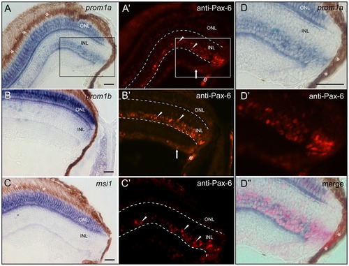
Co-expression analysis of zebrafish prominin-1a or b with Pax-6 and/or musashi-1 within retina cell layers. (A–D′′) Cryosections of eyes from adult zebrafish were processed for non-radioactive ISH using antisense DIG-labelled prominin-1a (A, D, D′′; prom1a), prominin-1b (B; prom1b) or musashi-1 (C; msi1) probe combined with IHC detection of Pax-6 (A′–C′, D′, D′′; anti-Pax-6). Arrowheads and arrows show the Pax-6 immunoreactivity (red) within the inner nuclear layer (INL) and ganglion cell layer (GCL), respectively. The area shown in the inset (A, A′) is displayed as zoom-in in panels D-D3. Note the expression of Pax-6 overlaps with prominin-1a within the INL. Asterisks indicate the retinal pigmented (dark brown) epithelium. Dashed lines delineate the INL. e, erythrocytes. Scale bars, 25 μm. |
| Genes: | |
|---|---|
| Antibody: | |
| Fish: | |
| Anatomical Terms: | |
| Stage: | Adult |