
Fig. S3
- ID
- ZDB-FIG-120201-36
- Publication
- Cheung et al., 2012 - Regulation of intrahepatic biliary duct morphogenesis by Claudin 15-like b
- Other Figures
- All Figure Page
- Back to All Figure Page
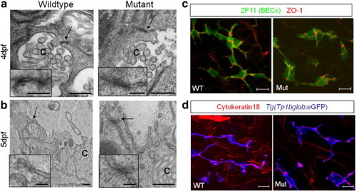
Junctions are present in cldn15lbfh290 mutant livers. Electron microscopy at 4 (a) and 5 (b) dpf. Insets are magnification of the arrowed areas showing junctional complexes that lie between hepatocytes and bile canaliculi [c]. Junctional complexes can be found in both wildtype and mutant livers. Scale bars are 0.4 μm (inset: 0.1 μm). (c) Whole-mount confocal projection of 110 hpf larvae immunostained with 2F11 and ZO-1(rabbit) to mark BECs and tight junctions between BECs, respectively. Tight junctions appear to be properly localized between BECs in the mutant livers. (d) Whole-mount confocal projection of 100 hpf Tg(Tp1bglob:eGFP)um14 larvae immunostained for cytokeratin-18 to mark the canals of Hering. These canals, which line the biliary ducts, appear normal in mutant livers. Cytokeratin-18 immunostaining also labels the intrahepatic vasculature. Scale bars in c and d are 10 μm. |
Reprinted from Developmental Biology, 361(1), Cheung, I.D., Bagnat, M., Ma, T.P., Datta, A., Evason, K., Moore, J.C., Lawson, N.D., Mostov, K.E., Moens, C.B., and Stainier, D.Y., Regulation of intrahepatic biliary duct morphogenesis by Claudin 15-like b, 68-78, Copyright (2012) with permission from Elsevier. Full text @ Dev. Biol.