
Fig. S3
- ID
- ZDB-FIG-120131-39
- Publication
- Wang et al., 2011 - Tfap2a and Foxd3 regulate early steps in the development of the neural crest progenitor population
- Other Figures
- All Figure Page
- Back to All Figure Page
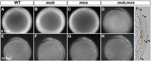
Apoptotic cells in the neural plate are increased in mob, mos, and mob;mos embryos. Lateral view (dorsal to the right) of wild-type (WT) (A, E), mob (B, F), mos (C, G), and mob;mos (D, H) live embryos stained with acridine orange dye at 8 (A–D) and 10 (E–H) hpf. At 8 hpf, only mob;mos embryos show a few dying cells in the neural plate, whereas at 10 hpf, dying cells are present in the neural plate of mob and mos embryos and dramatically increased in mob;mos embryos. (I) Coronal section of TUNEL-stained mob;mos embryos at 10 hpf. Arrows indicate TUNEL-positive cells at, or close to the neural plate surface. |
Reprinted from Developmental Biology, 360(1), Wang, W.D., Melville, D.B., Montero-Balaguer, M., Hatzopoulos, A.K., and Knapik, E.W., Tfap2a and Foxd3 regulate early steps in the development of the neural crest progenitor population, 173-85, Copyright (2011) with permission from Elsevier. Full text @ Dev. Biol.