
Fig. S4
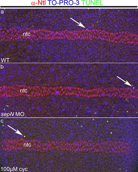
Loss of SepN does not affect cell death among adaxial cells. Cell death was measured in somitic mesoderm of three-somite stage WT (a) or sepN morphant (b) embryos. As a control to detect cell death that might occur whenever adaxial cell differentiation was blocked, embryos treated with 100 μM cyclopamine (c) were also examined. Adaxial cells form parallel rows of cells that lie immediately adjacent to the notochord and were visualized by a combination of staining for all nuclei (TO-PRO-3, blue) and for the No Tail protein, a marker of notochord (ntc) nuclei (α -Ntl, red). Apoptotic cells were visualized with TUNEL assay (green). Arrows indicate TUNEL-positive cells in the adaxial region, observed in similar numbers in control and treated embryos. Dorsal views with rostral left. |