
Fig. 3
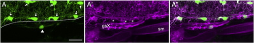
Sox:rfp+ peripheral glia contribute to the hindbrain transition zone of the Xth nerve. Images shown are from a 4 dpf olig2:gfp;sox:rfp larva (n = 3). Panel A is a dorsal view (anterior at left) of one side of the hindbrain, caudal to the otocyst, showing GFP+ oligodendrocytes and their processes. Arrows point to oligodendrocyte cell bodies and an arrowhead indicates an olig2-expressing cell present outside of the hindbrain. The dashed line marks the approximate position of the hindbrain boundary. A′ shows membrane-targeted RFP in the peripheral glia composing the vagal sheath, as well as in some CNS oligodendrocytes and their processes. Note the RFP+ processes (marked by asterisks) that appear to link the glial sheath to the hindbrain at several points. A" is a merge of A and A′. Notice that the GFP+ cell seen in panel A is present in the glial sheath. sm, skeletal muscle; gsX, glial sheath of the Xth nerve. Scale bars are 20 μm. |
| Genes: | |
|---|---|
| Fish: | |
| Anatomical Terms: | |
| Stage: | Day 4 |
Reprinted from Developmental Biology, 357(2), Cox, J.A., Lamora, A., Johnson, S.L., and Voigt, M.M., Diverse mechanisms for assembly of branchiomeric nerves, 305-17, Copyright (2011) with permission from Elsevier. Full text @ Dev. Biol.