
Fig. 1
- ID
- ZDB-FIG-110812-17
- Publication
- Liu et al., 2011 - Cell adhesion molecule cadherin-6 function in zebrafish cranial and lateral line ganglia development
- Other Figures
- All Figure Page
- Back to All Figure Page
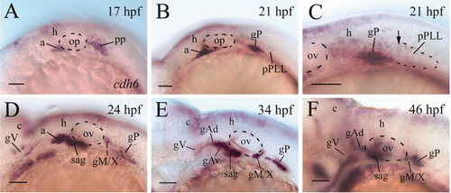
cdh6 expression in the cranial and lateral line ganglia of embryonic zebrafish. All panels show lateral views of the hindbrain region of whole-mount embryos (anterior to the left and dorsal up) processed for in situ hybridization using a cdh6 cRNA probe. Panel C is a higher magnification of the post otic area showing the newly formed primordium of the post lateral line (pPLL, dashed line indicating the boundary of its posterior 2/3). The arrow points to one of a few cdh6-expressing cells in the pPLL. a, anterior lateral line placode area; c, cerebellum; gAd, anterodorsal lateral line ganglion; gAv, anteroventral lateral line ganglion; gM/X, medial lateral line and vagal ganglia; gP, posterior lateral line ganglion; gV, trigeminal ganglion; h, hindbrain; op, otic placode; ov, otic vesicle (indicated by the dashed line); PP, postotic placode; sag, statoacoustic ganglion. Scale bar = 50 μm. |
| Gene: | |
|---|---|
| Fish: | |
| Anatomical Terms: | |
| Stage Range: | 14-19 somites to High-pec |