
Fig. 2
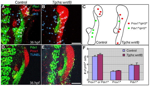
Wnt8a overexpression does not significantly affect cell proliferation or cell death in the endoderm 10 hours after heat-shock. Tg(hs:wnt8a) embryos were heat-shocked at 26 hpf and harvested at 36 hpf for whole-mount immunostaining. (A,B) Anti-Prox1 (red), anti-Pdx1 (green) and anti-pH3 (blue) triple labeling reveals that there was no significant difference in proliferation in Prox1+ or Pdx1+ endodermal cells between control (n=5 out of 5) and Wnt8a-overexpressing (n=4 out of 4) embryos 10 hours after heat-shock. Arrows point to Prox1+;pH3+ cells and arrowheads point to Pdx1+;pH3+ cells. (C) The distribution of pH3+ cells in A and B. Dotted lines outline the dorsal pancreas. (D,E) Anti-Prox1 (red), anti-Pdx1 (green) and TUNEL (blue) triple labeling reveals that no apoptosis was observed in Prox1+ or Pdx1+ endodermal cells of control or Wnt8-overexpressing embryos. (F) The number of pH3+ cells normalized over the size of the Prox1+ or Pdx1+ area are shown as a graph. Data are mean+s.e.m. Ventral views, anterior up. Scale bars: 50 μm. |