
Fig. 1
- ID
- ZDB-FIG-110622-55
- Publication
- Narayanan et al., 2011 - A transgenic wnt8a:PAC reporter reveals biphasic regulation of vertebrate mesoderm development
- Other Figures
- All Figure Page
- Back to All Figure Page
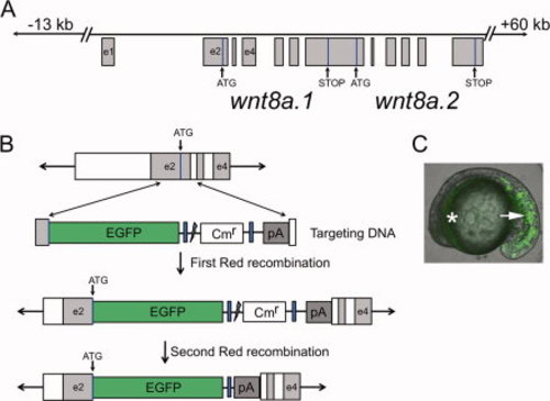
wnt8a gene structure and generation of an EGFP reporter PAC. A: Schematic diagram (not to scale) of the wnt8a locus within PAC clone 42J21. Line represents the PAC insert. Boxes indicate exons (e.g., e1). Start and stop codons are shown for both wnt8a coding sequences, although we targeted our insertion only into wnt8a.1. B: An outline of the homologous recombination procedure. Targeting DNA contains 50-bp ends homologous to exon 2 upstream of the translation start site and to the second intron (double arrows). Correct recombination results in an insertion of EGFP at the Wnt8a.1 translation start site. Blue boxes represent 50-bp repeated vector sequences that recombine during the second Red-recombination step that removes the chloramphenicol resistance gene (Cmr). This second Red recombination is stimulated by I-SceI cleavage at its target sequence, indicated by the jagged line. The EGFP coding sequence is followed by the SV40 poly adenylation signal (pA), then the rest of the wnt8a.1 locus. C: Transient EGFP expression from the modified PAC. Lateral view, anterior to the left. Note fluorescence in posterior mesoderm (arrow) and the YSL surrounding the yolk (asterisk). |