
Fig. s3
- ID
- ZDB-FIG-110622-119
- Publication
- Inoue et al., 2011 - One for all-a highly efficient and versatile method for fluorescent immunostaining in fish embryos
- Other Figures
- All Figure Page
- Back to All Figure Page
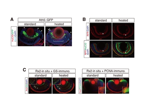
Application of heating method to co-staining with fluorescent proteins (FPs) in transgenic lines and whole-mount in situ hybridization. (A) HuC/D and pH 3 immunostainings of cryosections with Ath5::GFP medaka transgenic line. The heating method sufficiently retained GFP fluorescence after heating. GFP-positive retinal ganglion cells co-stained with HuC/D-positive retinal ganglion cells (a bracket) and pH 3-positive mitotic dividing cells (arrowheads). (B) HuC/D immunostaining of cryosections with Ath5::GAP43-RFP (RFP) zebrafish transgenic line. Note that fluorescent signal was still strong enough to detect after heating. (C) Whole mount in situ hybridization of medaka by uing antisense Rx2 probe in combination with either GS or PCNA immunostaining. Note that Rx2 co-stained with GS-positive Mueller glia cells (arrowheads) and PCNA-positive retinal progenitor cells at CMZ (brackets). Insets denote higher magnification of the overlap regions. Nuclei were counterstained with DAPI (blue). |