
Fig. 6
- ID
- ZDB-FIG-110516-42
- Publication
- Lazic et al., 2011 - Mef2cb regulates late myocardial cell addition from a second heart field-like population of progenitors in zebrafish
- Other Figures
- (all 15)
- All Figure Page
- Back to All Figure Page
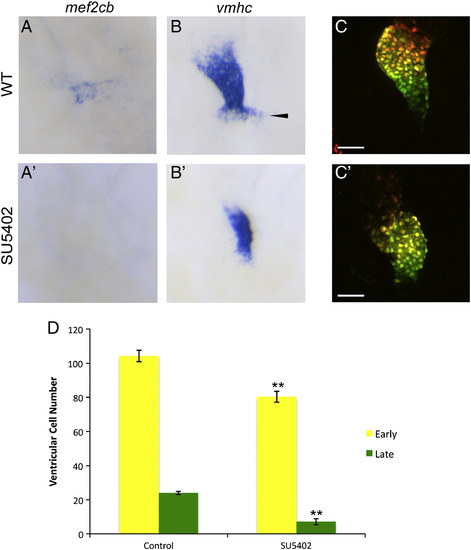
FGF signaling between 20 and 30 hpf was required for early and late myocardial addition. Chemical inhibition of FGF signaling between 20 and 30 hpf completely abolished mef2cb expression at 30 hpf (compare A to A′). At 30 hpf, the ventricle was smaller and the late ventricular region (arrowhead) was not seen in FGF inhibited embryos (compare B to B′). Photoconversion assay (D) between 30 and 48 hpf showed a smaller ventricle and less late myocardial addition in FGF inhibited embryos (C′) compared to DMSO treated control (C). Data shown as mean ± SEM; **P < 0.01. Scale bar represents 50 μm. |
| Genes: | |
|---|---|
| Fish: | |
| Condition: | |
| Anatomical Term: | |
| Stage: | Prim-15 |
| Fish: | |
|---|---|
| Condition: | |
| Observed In: | |
| Stage: | Prim-15 |
Reprinted from Developmental Biology, 354(1), Lazic, S., and Scott, I.C., Mef2cb regulates late myocardial cell addition from a second heart field-like population of progenitors in zebrafish, 123-133, Copyright (2011) with permission from Elsevier. Full text @ Dev. Biol.