Fig. S7
- ID
- ZDB-FIG-110516-28
- Publication
- Klein et al., 2011 - Neuron navigator 3a regulates liver organogenesis during zebrafish embryogenesis
- Other Figures
- All Figure Page
- Back to All Figure Page
|
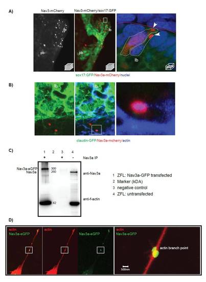
Nav3a protein is expressed in budding hepatoblasts in vivo and interacts physically with actin filaments. (A) Sox17:GFPs870 embryos were injected at the single cell stage with pDest-Tol2-sox17-nav3a-mCherry plasmid allowing imaging of endodermal Nav3a-mCherry expression in vivo. At 40 hpf embryos were fixed and analyzed for endodermal GFP and Nav3a-mCherry protein expression by confocal microscopy. Left: confocal stack showing Nav3a-mCherry protein in the budding liver. Middle: Merged view of the intestinal endoderm (GFP) and endodermal Nav3a-mCherry protein localization. Boxed area indicates the magnified area shown in the right picture. Right: Detailed view of Nav3a-mCherry protein localized in hepatoblast protrusions at the liver budding front (arrowheads). (B) Nav3a interacts in vivo with actin in budding endodermal cells. Claudin-GFP embryos express membrane-linked GFP in the endoderm at 30 hpf. Nav3a-mcherry was overexpressed by injection of pDest-Tol2-sox17-nav3a-mCherry plasmid. Embryos were fixed at 30 hpf and actin was detected by immunofluorescence with phalloidin-Alexa633 staining. (C,D) Nav3a interacts physically with f-actin and binds to actin branching points. ZFL cells were transfected with pDestTol2-CMV-nav3a-eGFP to induce overexpression of Nav3a-eGFP. Whole protein lysate was immune-precipitated with anti-Nav3a antibody. Precipitated protein was analyzed by Western blot for Nav3a and actin expression. As a Western Blot positive control unprecipitated protein lysate was used to verify the anti-Nav3a antibody and anti-actin antibody (lane 4). Lane 1 shows that in pTol-CMV-nav3a-eGFP transfected cells Nav3a-eGFP co-precipitates with actin, demonstrating binding of Nav3a-eGFP to f-actin. (D) Nav3a interaction with acting in spike like protrusions of filipodia extensions in Pac2 cells. A shows ventral view. lb, liver bud; pb, pancreatic bud. |

