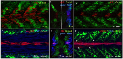
Activated Smads display a dynamic pattern of nuclear accumulation during zebrafish myotomal development. (A,B) Medial parasagittal optical and transverse sections of 14 ss embryos showing nuclear accumulation of pSmad (green) in slow myoblasts, marked by F59 (red) staining in A and prdm1a:GFP expression (blue) in B; the blue channel has been suppressed on the left-hand side of this panel to aid visualisation of the red and green signals. Note absence of accumulation in cells closest to the midline in both cases. These latter cells are the presumptive MPs. (C) 18 ss wild-type embryo showing the mutually exclusive pattern of Eng (4D9) and pSmad accumulation. (D-F) Lateral and medial parasagittal optical sections and transverse section of 20 ss Tg(eng2a:eGFP)i233 embryos. After the outward migration of slow fibres labelled with F59 (blue) in D, F, and prdm1:GFP (blue) in E, pSmad accumulates in a subset of medially located fast fibres, except for those at the midline, closest to the notochord that express the transgene (red in D,F) and endogenous Eng, revealed by 4D9 (red in E). Note that at this stage, most eng expression is still restricted to MPs, but is starting to initiate in MFFs in more anterior somites (arrows in F).
|

