
Fig. 9
- ID
- ZDB-FIG-101207-10
- Publication
- Choi et al., 2010 - In vivo development of dendritic orientation in wild-type and mislocalized retinal ganglion cells
- Other Figures
- All Figure Page
- Back to All Figure Page
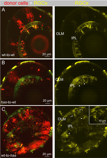
Dendritic misprojections of ectopically located retinal ganglion cells in the has mutant is non-cell autonomous. In each chimera, donor cells were labeled by a lineage tracer (red), fluororuby. Donor cells were always taken from the isl2b:MGFP line (yellow), either in a wild-type (wt) or has background, to better visualize dendrites. (A) Wild-type donor cells transplanted to a wild-type host. RGC dendritic arbors were oriented toward the OLM and stratified within an IPL. (B) has donor cells transplanted to a wild-type host. All has RGCs were located in the GCL (arrowheads). C. Wild-type donor cells transplanted to a has host. Most RGCs were located in the GCL (arrowheads), but a few cells were located ectopically (arrows). Inset shows higher magnification of the misplaced RGCs indicated by the arrows. |