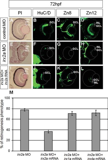
irx2a is required for neurogenesis of the retina ganglion cells. A–L: Immunostaining of 72-hpf retina. A–I: Lamination defects of irx2a MO2 morphant retinas were revealed by H&E staining. In the control morphant (A), the retina was segregated into three distinct nuclear layers (outer nuclear layer [ONL], inner nuclear layer [INL, and ganglion cell layer [GCL]). In contrast, the laminar structure was disrupted in the irx2a MO2 morphant retina (E). HuC/D antibody was used to label differentiated RGCs in the GCL (B). However, HuC/D staining was barely detectable in the morphant retina (F). Zn8 immunoreactivity was observed in the surface of RGC and axons in the optic nerve (C). Zn8 staining was completely absent in the morphant (G). Zn12 immunostaining labeled RGCs and amacrine cells in the INL of the control retina (D), but only a small patch of staining was detected in the morphant retina (H). Neurogenesis defects in the GCL and INL of the morphant retina were rescued by injection of irx2a mRNA (I–L). M: Co-injection of irx2a mRNA, but not irx1a, irx4a, rescued the irx2a morphant phenotype.
|

