
Fig. S2
- ID
- ZDB-FIG-100913-12
- Publication
- May-Simera et al., 2010 - Bbs8, together with the planar cell polarity protein Vangl2, is required to establish left-right asymmetry in zebrafish
- Other Figures
- All Figure Page
- Back to All Figure Page
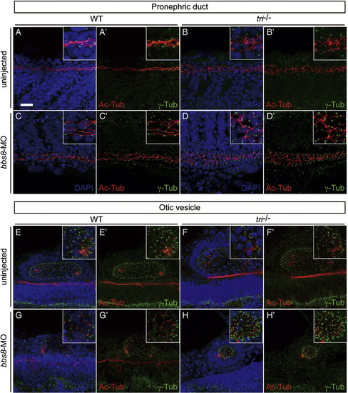
Cilium/basal body structure in the pronephric duct and otic vesicle is disrupted in tri mutant. Cilia (Ac-Tub, red) and basal bodies (γ-Tub, green) were observed in the pronephric duct (A-D, A′-D′) and otic vesicle (E-H, E′-H′), counterstained with (A-H) or without (A′-H′) nuclei (DAPI, blue). The normally linear organisation of pronephric duct cilia in control siblings (A, A′ insets) is disordered in bbs8-morphants and tri-/- with notable dilation of the pronephric lumen (C, C′ insets and B, B′ insets). These affects are further exacerbated in tri-/- embryos injected with bbs8-MO (D, D′ insets). Similarly, in otic vesicles (E-G insets, E′-G′ insets), the classical oblong shape in control sibling embryos (E, E′ insets) is malformed to a more oval shape in tri-/- (F, F′, insets) and an undersized sphere in bbs8-morphants (G, G′ insets). Again these defects are enhanced in tri-/- embryos injected with bbs8-MO, generating even smaller spherical vesicles with notable compaction of cilia/basal bodies (H, H′ insets). Taken together, these data suggest vangl2/tri and bbs8 are required for normal cilium/basal body structure in a variety of tissues. Scale bar: 200 μm. |
Reprinted from Developmental Biology, 345(2), May-Simera, H.L., Kai, M., Hernandez, V., Osborn, D.P., Tada, M., and Beales, P.L., Bbs8, together with the planar cell polarity protein Vangl2, is required to establish left-right asymmetry in zebrafish, 215-225, Copyright (2010) with permission from Elsevier. Full text @ Dev. Biol.