
Fig. S4
- ID
- ZDB-FIG-100806-23
- Publication
- Talbot et al., 2010 - hand2 and Dlx genes specify dorsal, intermediate and ventral domains within zebrafish pharyngeal arches
- Other Figures
- All Figure Page
- Back to All Figure Page
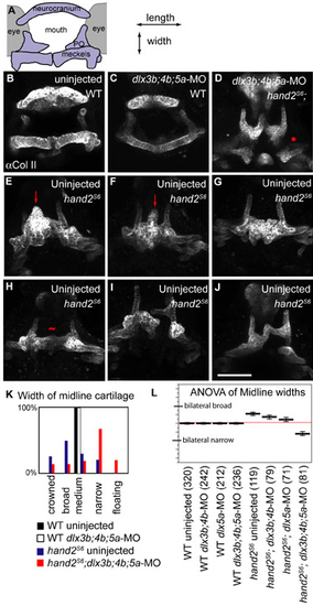
The shapes of first arch-derived skeleton are highly variable in hand2S6, but show significant changes after dlxb3;4b;5a-MO injection. (A) Schematic of anterior view, medial towards the center, dorsal upwards. (B-J) Anterior views of fish immunolabeled for Collagen IIa1. E is the same fish as is shown in Fig. 8C, for comparison of lateral view with anterior view. Images are projections of confocal stacks. (B) Uninjected and (C) dlx3b;4b;5a-MO injected wild-type fish have similar ventral shapes. (D) hand2S6 homozygotes injected with dlx3b;4b;5a-MO typically have narrowed midline cartilages (narrow), which may be separated from posterior PQ (asterisk: floating). (E-J) A phenotypic series of midline cartilage shapes in uninjected hand2S6. (E,F) The ventral midline sometimes contains a cartilaginous peak (arrow: crowned). (G) In other fish, the palatoquadrate expands (broad), but without ectopic pterygoids. (H) The ventral cartilages are sometimes similar to wild type in width (tilde: medium), though they are shortened in length. (I) Cartilages are not always bilaterally symmetrical. (I,J) Rarely, narrow cartilages are also seen in uninjected hand2S6; (K) however, a histogram of fish in each shape class shows that uninjected hand2S6 homozygotes typically look dramatically different from hand2S6 homozygotes injected with dlx3b;4b;5a-MO. (L) The shape classes were converted into a numeric score of midline size, and the means of these scores are plotted as thick black lines. Global mean is indicated with a thin red line. Error bars are 95% confidence intervals, determined by ANOVA. Number of fish scored is given in parentheses. Scale bar: 100 μm. |