
FIGURE
Fig. 3
- ID
- ZDB-FIG-100628-64
- Publication
- Kim et al., 2010 - Gli2a protein localization reveals a role for Iguana/DZIP1 in primary ciliogenesis and a dependence of Hedgehog signal transduction on primary cilia in the zebrafish
- Other Figures
- All Figure Page
- Back to All Figure Page
Fig. 3
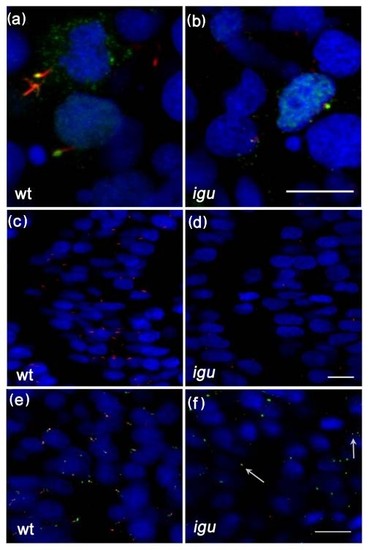
Primary cilia are truncated or missing from somitic cells in Iguana (igu) mutant embryos. Gli2a-GFP co-localized with acetylated tubulin (red) in igu mutant embryos in presumptive axonemes (b) that were severely truncated relative to those of wild type primary cilia (a). Truncated cilia (red) were only occasionally present in igu embryos (d) in contrast to wild type (c). Basal bodies (green: γ-tubulin in e, f) were present in all cells in igu mutant embryos (f) as in wild type (e) and were associated with the truncated axonemes (arrows). Scale bars: 10 μm |
Expression Data
Expression Detail
Antibody Labeling
Phenotype Data
Phenotype Detail
Acknowledgments
This image is the copyrighted work of the attributed author or publisher, and
ZFIN has permission only to display this image to its users.
Additional permissions should be obtained from the applicable author or publisher of the image.
Open Access.
Full text @ BMC Biol.