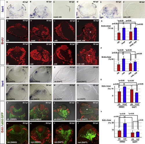
Neurogenic defects in the ncad and nok mutant retinas depend on the Notch signaling pathway. (A–F) Expression of her4 mRNA in 48 hpf retinas of ncad (B), ncad; mib (C), nok (E), and nok; mib (F) mutants and their wild-type siblings (A and D). her4 mRNA is expressed in the central part of CMZ of wild-type retinas (A and D, lines and arrows). On the other hand, a patchy pattern of her4 expression is observed in the central region in ncad (B) and nok mutant retinas (E). her4 mRNA expression level is markedly reduced in the ncad; mib (C) and nok; mib mutant retinas (F). (G–J) BrdU labeling of wild-type retina (G) and ncad (H), mib (I), and ncad; mib mutant retinas (J) at 50 hpf. BrdU incorporation is observed in CMZ of the wild type and mib mutant retinas, whereas the central retina is BrdU-positive in the ncad and ncad; mib mutants, suggesting that mib mutation does not efficiently suppress BrdU incorporation in the central retina of ncad mutants (J, asterisk). (K) Ratio of number of BrdU-positive cells to total number of retinal cells in wild-type sibling retina and ncad, mib, and ncad; mib mutant retinas. The higher level of BrdU incorporation in the ncad mutant retina is not decreased by mib mutation. (L–O) BrdU labeling of wild-type retina (L) and nok (M), mib (N), and nok; mib mutant retinas (O) at 56 hpf. BrdU incorporation is observed in CMZ of the wild type and mib mutant retinas, whereas the central retina is BrdU-positive in the nok and nok; mib mutants (O, asterisk). (P) Ratio of number of BrdU-positive cells to total number of retinal cells in wild-type sibling retinas and nok, mib, and nok; mib mutant retinas. The higher level of BrdU incorporation in the nok mutant retina is partially decreased by mib mutation. (Q–T) her4 mRNA expression in wild-type sibling retina (Q) and ncad mutant retina (R) treated with DMSO, and in wild-type sibling retina (S) and ncad mutant retina (T) treated with DAPT at 58 hpf. At this stage, her4 mRNA expression level decreases in the central retina and the expression is observed only in CMZ of the ncad mutant retina (R). her4 mRNA expression is completely absent in both the wild-type retina and ncad mutant retina treated with DAPT (S and T). (U–X) her4 mRNA expression in wild-type sibling retina (U) and nok mutant retina (V) treated with DMSO, and in wild-type sibling retina (W) and nok mutant retina (X) treated with DAPT at 58 hpf. In contrast to the ncad mutant, her4 mRNA expression is observed in the central retina as well as CMZ of the nok mutant (V). her4 mRNA expression is completely absent in both wild-type and nok mutant retinas treated with DAPT (W and X). (Y and Z, a and b) ath5:GFP expression (green) and EdU incorporation (red) in wild-type sibling retina (Y) and ncad mutant retina (Z) treated with DMSO, and in wild-type sibling retina (a) and ncad mutant retina (b) treated with DAPT at 58 hpf. EdU incorporation is suppressed but ath5:GFP expression is observed in the central retina of the ncad mutant treated with DAPT (asterisk in b). (c–f) ath5:GFP expression (green) and EdU incorporation (red) in wild-type sibling retina (c) and nok mutant retina (d) treated with DMSO, and in wild-type sibling retina (e) and nok mutant retina (f) treated with DAPT at 58 hpf. EdU incorporation is suppressed but ath5:GFP expression is observed in the central retina of the nok mutant treated with DAPT (asterisk in f). (g) Ratio of number of EdU-positive cells to total number of retinal cells in wild-type sibling retina and ncad mutant retinas treated with DAPT or DMSO. The higher level of EdU incorporation in the ncad mutant retina was decreased by DAPT treatment. (h) Ratio of number of EdU-positive cells to total number of retinal cells in wild-type sibling retina and nok mutant retinas treated with DAPT or DMSO. The higher level of EdU incorporation in the nok mutant retina is decreased by DAPT treatment. Statistical analyses were carried out by Student’s t-test (K and P, g and h).
|

