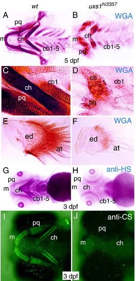
Proteoglycan detection in wild-type and uxs1 mutant skeletons. (A–F) Whole-mount wheat germ agglutinin (WGA) staining to visualize N-acetylglucosamine, ventral views. (G–J) Whole-mount immunostaining against heparan sulfate (G, H) and chondroitin sulfate (I, J) proteoglycans, ventral views. Dissected pharyngeal cartilages revealed reduced WGA staining in uxs1hi3357 mutants (B, D), compared to wild-type siblings (A, C) at 5 dpf. Higher magnification of ceratohyal regions also showed that WGA-positive material was not deposited normally in mutants (D), compared to organized deposition in wild types (C). Dissected pectoral fins showed that both endoskeletal disc and actinotrichia had less WGA staining and fewer actinotrichia in uxs1hi3357 mutants (F), compared to wild-type siblings (E) at 5 dpf. Immunodetection of heparan sulfate demonstrated that HSPGs were localized to pharyngeal domains in wild type (G), but HSPGs were not detectable in homozygous uxs1hi3357 animals (H). Similarly, immunodetection of chondroitin sulfate was abundant in wild-type cartilages (I), but was absent in uxs1 mutants (J). Abbreviations: at, actinotrichia; cb1-5, ceratobranchials 1-5; ch, ceratohyal; ed, endoskeletal disc; m, Meckel′s cartilage; pq, palatoquadrate.
|
