
Fig. 1
- ID
- ZDB-FIG-100114-25
- Publication
- Codina et al., 2010 - Loss of Smyhc1 or Hsp90alpha1 function results in different effects on myofibril organization in skeletal muscles of zebrafish embryos
- Other Figures
- All Figure Page
- Back to All Figure Page
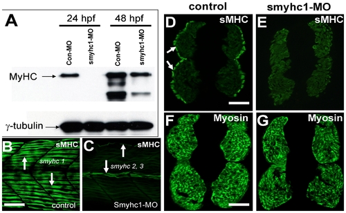
Knockdown of smyhc1 expression by smyhc1 ATG-MO. A. Western blot analysis shows the effect of smyhc1-MO on the expression of the myosin heavy chain in slow muscles (F59) at 24 and 48 hpf. Anti-γ-tubulin was used as a loading control. B, C. Anti-MyHC antibody (F59) staining shows myosin expression in trunk slow muscles of control (B), or smyhc1 knockdown (C) embryos at 48 hpf. Myosin expression was significantly knocked down in slow muscles. However, myosin expression could be detected in myofibers in the dorsal and myoseptum region of the myotome (arrows) that express smyhc2 and smyhc3. D, E. F59 antibody staining on cross-sections shows MyHC expression in slow muscles (arrows) of control (D) or smyhc1-ATG-MO injected embryos (E) at 48 hpf. F, G. MF20 antibody staining shows MyHC expression in fast muscles of control (F) or smyhc1-ATG-MO (G) injected embryos at 48 hpf. Scale bars = 25 μm in B; 75 μm in D and F. |
| Antibody: | |
|---|---|
| Fish: | |
| Knockdown Reagent: | |
| Anatomical Terms: | |
| Stage Range: | Prim-5 to Long-pec |